
25+
Years of Excellence
1,00,000+ Successful Surgeries
20+
Advanced Technologies
Expert Refractive
Surgeons
Advanced wavefront-guided LASIK technology provides highly personalized vision correction by mapping your unique eye characteristics for sharper, high-quality vision.
TransPRK is a completely touch-free, flap-free laser vision correction procedure that offers safe treatment with minimal discomfort and a smooth recovery.
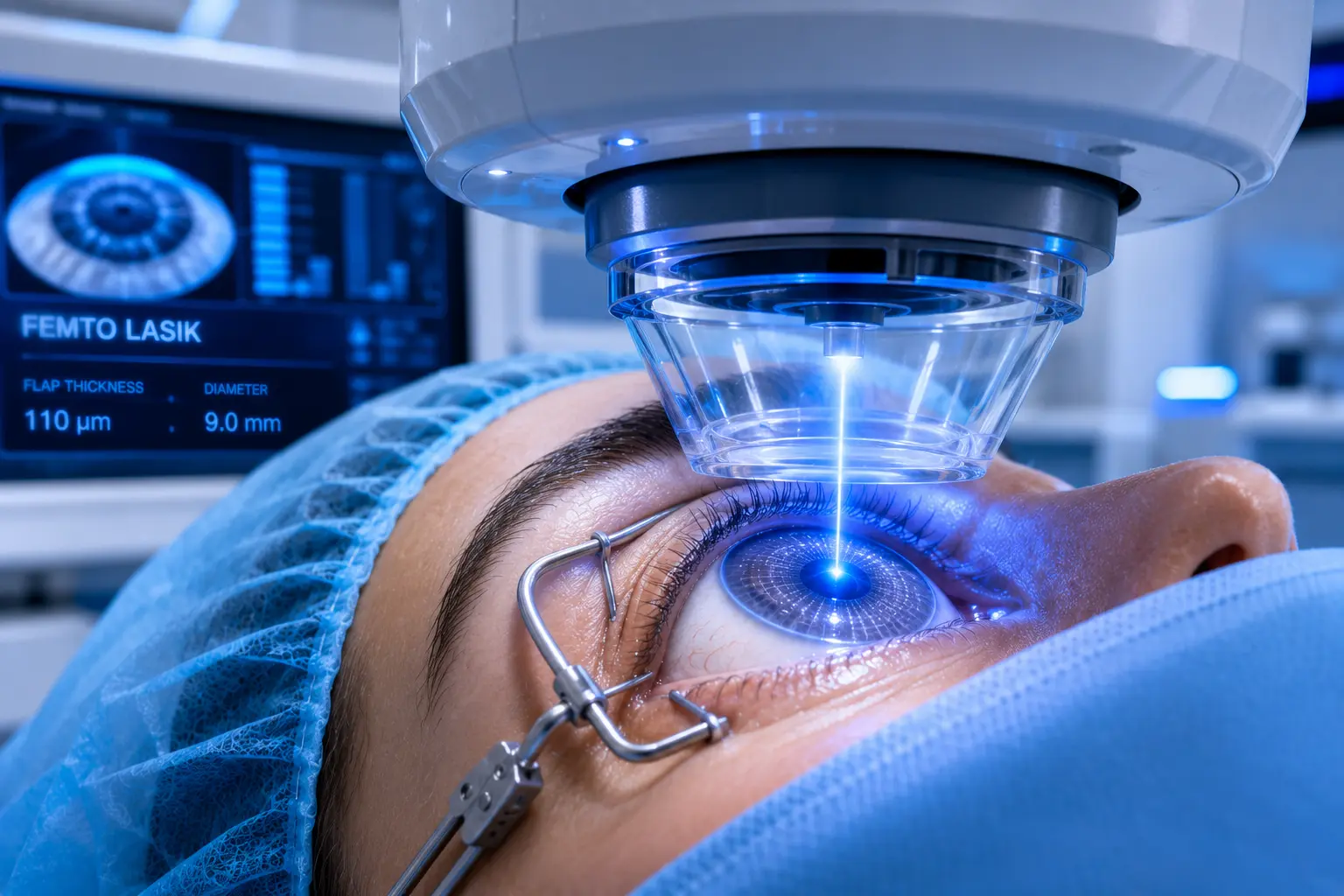
Femto LASIK uses advanced femtosecond laser technology to create a precise corneal flap, providing accurate, blade-free vision correction with faster recovery.
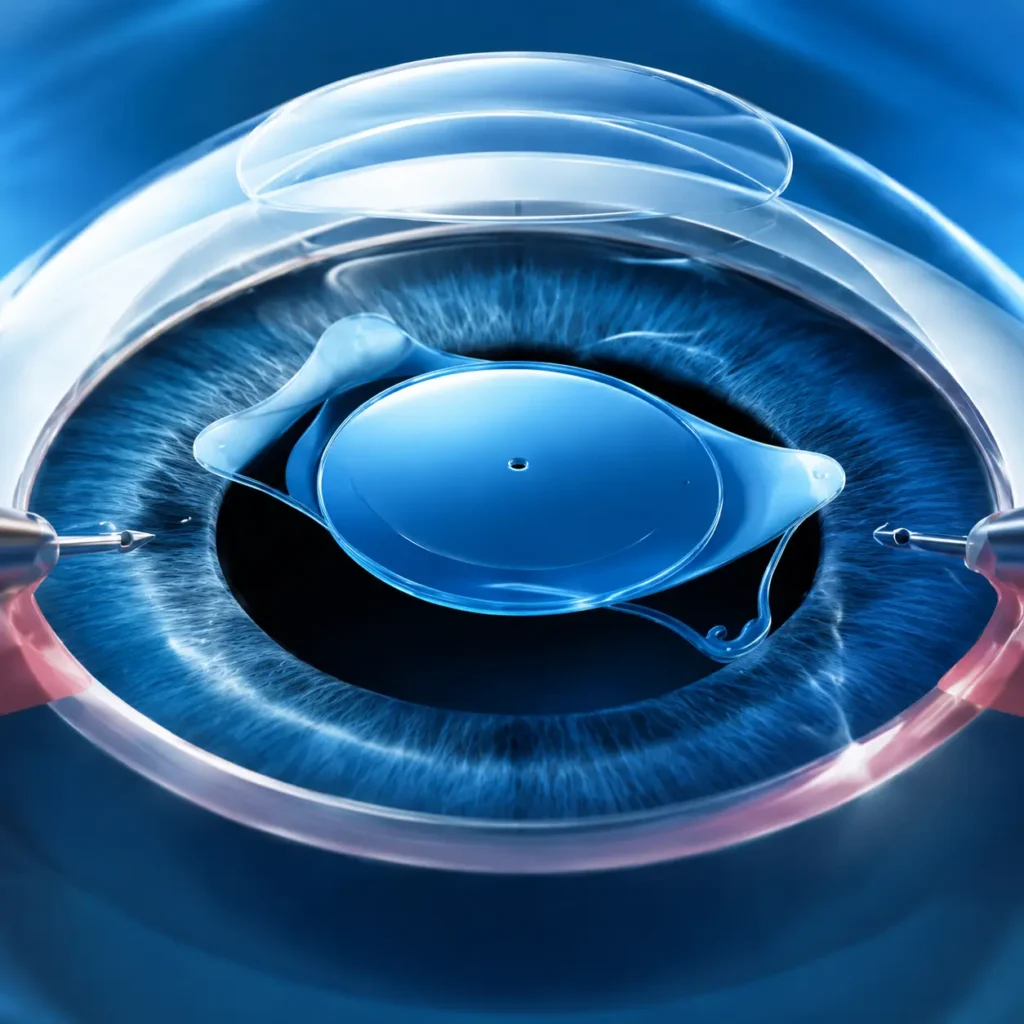
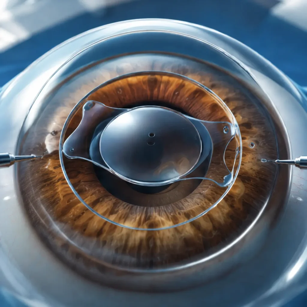
ICL (Implantable Collamer Lens) is an implantable lens procedure for patients with high refractive errors or thin corneas who are not suitable candidates for LASIK.
IPCL is an implantable phakic contact lens procedure designed to correct high myopia, hyperopia, and astigmatism without removing the natural lens.
PresbyLASIK is an advanced laser vision correction procedure designed to improve both near and distance vision for people experiencing age-related reading vision problems (presbyopia).
State-of-the-art laser systems
Personalized treatment plans tailored to your vision needs.
Experienced refractive surgeons specializing in LASIK, SMILE, PRK, and ICL procedures.
Safe, minimally invasive treatment with faster recovery.
Trusted by thousands of satisfied patients with proven clinical outcomes.
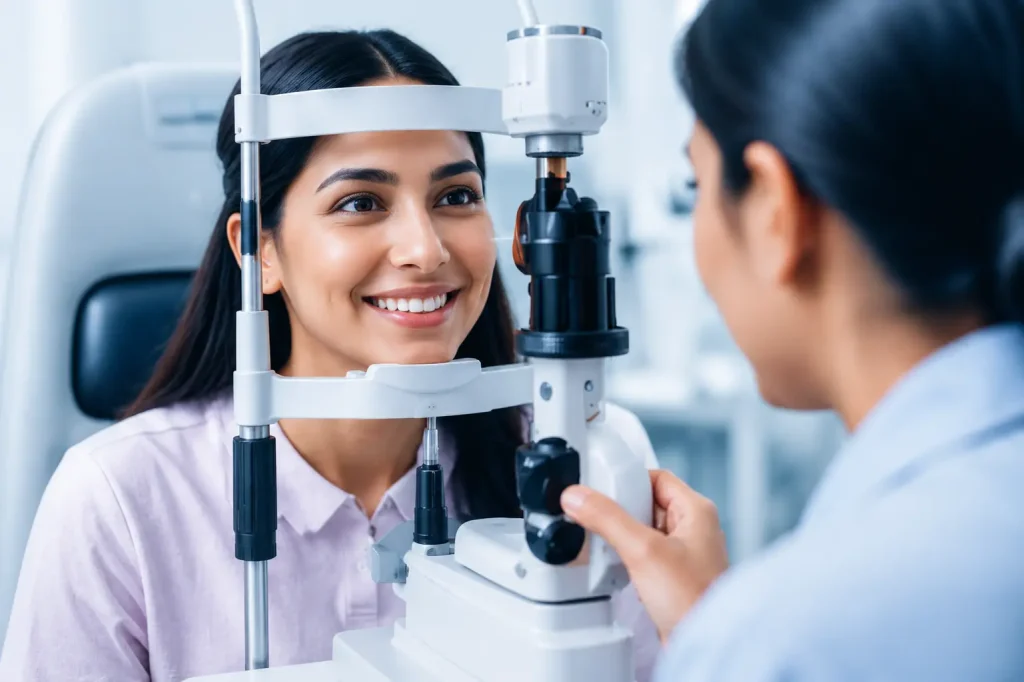
Schedule a comprehensive eye evaluation with our experienced refractive specialists to determine the most suitable vision correction procedure for your eyes and lifestyle
Advanced Eye Care Technology
Our advanced diagnostic systems, wavefront-guided technology, and precision laser platforms help create personalized treatment plans for safer procedures, faster recovery, and excellent visual outcomes.